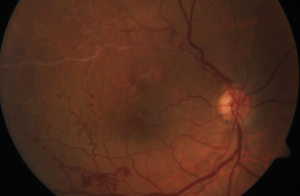
Laguna Hills Retinal Vein Occlusion
The retina is the light-sensing layer of cells that lines the inner wall of the back of the eye. The retina converts light into signals that are sent to the brain where they are recognized as images.
What is Retinal Vein Occlusion?
Your retinas require a rich supply of oxygenated blood. This blood is carried to the retina from the heart through arteries, while veins carry deoxygenated blood from the retina back to the heart via the optic nerves. The arteries that transport oxygen to your retina may stiffen and thicken over the years, especially in people with diabetes, cardiovascular disease, high blood pressure and high cholesterol. The retinal arteries and veins are close to the optic nerve and cross various locations on the retina’s surface. As arteries thicken, they can compress the retinal vein at artery-vein crossing points or in the optic nerve. This compression can be so severe that the veins become constricted and thus blood flow to the heart is reduced. The blood then backs up into the retina not unlike the water in a clogged bathtub. The pressure of blood in the veins causes swelling (called macular edema) and bleeding in the retina. There are three kinds of retinal vein occlusions:
- The most commonly seen cases are branch retinal vein occlusions, in which the blockage occurs in a smaller vein on the retina’s surface. The consequent hemorrhaging and swelling may result in diminished vision if the macula, the retina’s center, is involved.
- The second most common form of retinal vein occlusion is a central retinal vein occlusion in which the main retinal vein is blocked within the optic nerve. This is usually a more severe kind of occlusion that causes hemorrhages and swelling throughout the retina. This often leads to a more serious decrease in sight.
- Less common is hemi-retinal vein occlusion, in which the vein draining one-half of the venous circulation is occluded. Generally, this is more severe than the branch retinal vein occlusion but less so than central retinal vein occlusion
Eyes with retinal venous occlusive disease may develop macular edema (retinal swelling), or growths of fragile, abnormal blood vessels on the retina’s surface. These growths can cause bleeding into the vitreous gel. This bleeding is typically associated with the sudden appearance of floaters or severe loss of vision. Patients with central retinal vein occlusions may also develop abnormal blood vessels on the iris. These blood vessels can cause a severe form of glaucoma known as neovascular glaucoma.
Fortunately, retinal vein occlusion is usually only present in one eye so the other eye remains unaffected.
What are the risk factors for Laguna Hills retinal vein occlusions?
The main risk factors include: high blood pressure, diabetes, high cholesterol, cardiovascular disease, glaucoma, and other blood disorders. Some patients have no risk factors and a more extensive work-up to determine underlying cause may be warranted.
Symptoms of Laguna Hills Retinal Vein Occlusion
People with a retinal vein occlusion may not exhibit symptoms but those who do may have a sudden onset of blurred vision in one eye. However, blurred vision is a symptom of many ocular conditions. Therefore, one will have to undergo testing to confirm that one has retinal vein occlusion.
Treatment for Laguna Hills Retinal Vein Occlusion
There are numerous treatment options to reduce the risk of vision loss in cases of retinal vein occlusion. For those with macular edema resulting from this condition, laser photocoagulation may help. Anti-vasccular endothelial growth factors (anti-VEGF) drugs such as ranibizumab (Lucentis), bevacizumab (Avastin) or aflibercept (Eylea) may be injected into the vitreous gel to treat the swelling associated with this condition.
Like many medical conditions, the treatment is done to manage the disease and reduce the risk of worsening. Thus, with intravitreal injections, patients may require life-long treatment.
Retina specialists can treat patients with the more severe kind of venous occlusive disease (neovascular glaucoma) with scatter laser to the retina and/or continued injections in hopes of stabilizing the disease process and preventing severe vision loss or blindness.
As retinal vein occlusions are typically associated with high cholesterol, diabetes and high blood pressure, monitoring and treating these medical conditions are important in those patients with venous occlusive disease. Thus, following up with your primary care doctor is imperative.
Contact Our Laguna Hills Eye Doctors for Help with Retinal Vein Occlusion or Macular Edema
To learn more about venous occlusive disease and how our retina specialists can help, contact our physicians at Retina Associates of Orange County.

