

Retinal Tears & Detachment
What Is The Retina?
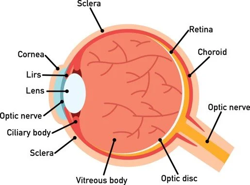
The retina is a thin sheet of nerve cells that lines the inside back wall of the eye. If the eye were a camera, the retina would be the film of that camera. Light travels through the eye and ultimately reaches photoreceptors, the light-sensitive cells in the retina. The signal from the photoreceptors then travel along the optic nerve to the brain, where the signals are interpreted as vision.
What Is The Vitreous?
The inside of the eye is filled with a gel called the “vitreous.” At birth, the vitreous is a formed gel and firmly adherent to the retina.
What Is A Posterior Vitreous Detachment (PVD)?
With age, the vitreous gel shrinks and becomes increasingly liquified. Eventually, the back edge of the vitreous pulls away from the retina to which it was previously adherent. This separation event is called a “posterior vitreous detachment” (PVD). PVD is a normal process of aging and occurs in most everyone, eventually (usually in one’s 50s or thereafter). Some patients experience no symptoms from their PVD, while others may notice flashes, floaters, or decreased vision.

If you have new floaters, new flashes, a curtain in the vision, or decreased vision, a prompt dilated retinal examination is required to evaluate for potential vision-threatening complications of the PVD process.
New or worsening floaters may be a sign of the following:
-
Posterior Vitreous Detachment
-
Vitreous Hemorrhage
-
Retinal Tear
-
Retinal Detachment
-
Inflammation
-
Benign Age-Related Changes
The only way to distinguish between the above entities is to undergo a careful, dilated retinal examination by an experienced eye doctor.
Problems That Can Occur During The PVD Process
As the vitreous pulls away from the retina, it may be unusually adherent in some areas, tugging on the retina and causing problems such as:
-
Vitreous Hemorrhage
If a blood vessel is tugged on, some bleeding may occur, which can cause lots of floaters or blurry vision or loss of vision.
-
Retinal Tears
As the vitreous gel tugs on the retina, it can tug hard enough to tear the retina (“retinal tear”). Up to 14% of patients with symptoms from a PVD may have a retinal tear. While the risk is highest at the first visit, there is an increased risk of new tears developing subsequently, especially the first 6-8 weeks after the PVD symptoms began.
-
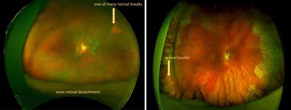
Retinal Detachment
Without treatment, up to 50% of retinal tears result in retinal detachment. A retinal detachment occurs because fluid enters through the retinal tear, allowing the retina to detach from the eye wall. As an analogy, consider a tear in the wallpaper of the bathroom. If the tear is left unfixed, steam from the shower will eventually cause the entire wallpaper to start peeling off.
Retinal detachment results in vision loss, initially in the peripheral vision and then progressing to involve the central vision.

Retinal Tear & Detachment Treatment
Retinal detachment is treatable when caught early. Treatment may or may not involve an in or outpatient surgical procedure. In cases that do require surgery, a patient may require immediate attention or the procedure may be postponed for days or even weeks depending on the condition’s severity.
Treatment Of A Retinal Tear
Treatment of a retinal tear reduces (but does not eliminate) the risk of progression to retinal detachment. Treatment involves laser (or less commonly, cryotherapy). The laser treatment is applied in the office, with no need for anesthesia other than topical drops, by special equipment that shines a light into the eye (similar to what happens during the eye exam) and then allows placement of laser spots in the area of concern
Laser treatment seals the retina surrounding a tear to the eyewall, such that the retina cannot easily detach in that area (like tacking down the retina surround the tear). Laser does not eliminate the tear nor does it eliminate or change symptoms of flashes or floaters. Laser reduces but does not eliminate risk of the tear progressing to retinal detachment. Laser also does not eliminate the risk of future tears occurring elsewhere in the retina.
Most patients can return to their usual activities after laser is performed, and close follow-up is performed for the weeks that follow to monitor for progression to retinal detachment, new tears, etc.
Treatment Of A Retinal Detachment
Retinal detachments may be treated in one of several ways, depending upon the specific anatomy and other considerations.
- Pars Plana Vitrectomy
Pars Plan Vitrectomy is a micro-incisional surgery whereby the vitreous pulling on the retina is removed, the fluid is drained from under the retina, the retina is reattached, and then the tears are treated with laser.
The laser is like glue such that it takes time to “dry.” Thus, either a gas bubble or an oil bubble is placed in the eye to hold the retina in place while the laser “dries.” Patients cannot see well through the gas (and to some extent, also the oil bubble). The gas resolves slowly on its own over 3-8 weeks. Oil required a second surgery for removal 3 or more months later.
Usually, the patient will need to position the head in a specific way (for example, “face down”) for 5-10 days to ensure the gas or oil bubble is supporting the retinal breaks.
Doctors may recommend this surgery in instances where:
- Diabetic retinopathy led to the retinal detachment
- Hemorrhage or other opaque substances have prevented a clear view into the eye
- The retinal tears are very far in the back of the eye
- Evidence of scarring appears on the retina’s surface
- Other conditions are affiliated with retinal detachment
- There are complications with cataract surgery
- Inflammatory disease occurs

- Scleral Buckle
A scleral buckle operation can be performed independently or in addition to a vitrectomy. In this procedure, your doctor places a small silicone band or implant around part or the entire eye, creating an indent in its wall, counterbalancing the force that caused the retinal tear and retinal detachment. Often, surgeons will drain the fluid under the detached retina, allowing it to settle more quickly into its natural position. An air gas bubble may or may not be placed in the eye for further support of retinal breaks. Usually, surgeons perform this procedure with general anesthesia, but local anesthesia may be used. Scleral buckle is typically an outpatient surgery.

- Combination Surgery
In some cases, the doctor might perform both a pars plan vitrectomy and scleral buckle surgery in order to treat a detachment.
- Pneumatic Retinopexy
In some cases, the retinal detachment can be repaired by injecting a gas bubble in the eye in the office. With diligent head positioning, the gas bubble can “plug” the retinal break and allow the retina to reattach. Laser can then subsequently be applied to seal the retinal break.
- Laser Barricade
The retina is not reattached but rather a barrier of laser is placed to tack the retina down at the edge and reduce risk of progression.

Contact Our Eye Doctors Today
To learn more about retinal detachment and its treatment options, contact our office at Retina Associates of Orange County. The sooner you speak to a retinal specialist, the sooner you can save and restore your sight.
Retinal Tear & Detachment Treatment FAQs
If you or a loved one has suffered from a retinal detachment or retinal tear, you may be wondering what the implications are and how this condition can be treated. Retinal detachment is a serious condition that often needs to be treated in a timely manner.
What Is A Retinal Tear?
Occasionally when the vitreous gel separates from the retina, it will pull on the retina, causing a retinal tear. Retinal tears can lead to a retinal detachment if fluid passes under the tear and separates the retina from the wall of the eye. Retinal tears can often be treated in the office with laser or cryotherapy (freezing).
What Is Retinal Detachment?
A healthy retina is attached to the inside wall of the eye. Detachment occurs when the retina is torn away from this position. The detached portion of the retina cannot see light well, resulting in blurred or even lost vision. Retinal detachments can progress rapidly, causing complete blindness if not fixed in time. Luckily there are multiple ways to repair a detached retina to reduce the risk of further vision loss.
What Are The Symptoms Of A Retinal Tear Or Retinal Detachment?
The classic symptoms for a retinal tear are similar to those of the normal vitreous gel separation and include flashes and floaters.
Flashes and/or floaters may also be symptoms of retinal detachment, but detachment usually also entails decreased vision or a visual field darkening in the peripheral vision—like a shadow or shade coming across the eye.

How a Retinal Tear or Detachment Affects Your Vision
The retina is one of the most essential parts of the eye. A damaged retina can severely limit your vision, which can lead to permanent health consequences. Treating any type of retinal issue is critical if you want to improve your functional vision and maintain the health of your eyes. Treatment will significantly hinder the advancement of your condition so that it does not get worse. If you believe you are suffering from a retinal tear or detachment, seek immediate treatment right away by starting with a consultation with an experienced retina specialist.
Recognizing Common Retinal Issues
If you are suffering from a retinal tear, you may have difficulty doing everyday tasks and basic activities like driving, reading, preparing meals, or watching television. Additionally, constantly having to blink and rub your eyes because of floaters can indicate eye issues.
A retinal detachment is more noticeable and severe. This occurs when the retina is completely separated from the rest of the eye, resulting in severely compromised vision. If it is not immediately treated, it can advance quickly and can lead to permanently affected vision. Surgical procedures can treat retinal detachments so that vision is restored. Eye surgery must be done in the early stages of a retinal detachment to have the highest chances of success. Talk to a trusted eye doctor who has a record of successful surgeries for patients with retinal tears or detachments as soon as possible.
Why See an Eye Doctor
It could be time to see a Laguna Hills eye doctor for retinal tear and retinal detachment if you believe that you have symptoms of eye issues. There are several procedures and treatments available to help restore your vision and prevent a complete loss of vision. Seeing an eye doctor as soon as you notice potential signs of eye problems is essential so that you can receive the proper treatment. The earlier you decide to see a doctor, the higher your chance of improving your vision.
What To Expect From Your Eye Exam
As a skilled and experienced Laguna Hills eye doctor retinal tear and retinal detachment specialist can explain to you, there are several things that you can expect from a comprehensive diagnostic exam. The doctor will ask you what kind of symptoms you have been experiencing and for how long. They will ask about your medical history, family history and lifestyle choices that could impact your health. During the exam, the doctor will evaluate the health of your eyes with several diagnostic tools and instruments, as well as have you do a vision test and report the ways in which your vision is obscured.
After Receiving Treatment
You may feel soreness and discomfort after undergoing outpatient eye surgery to treat retinal detachments. You can expect your recovery period to take anywhere from weeks to several months. Always follow the doctor’s orders for post-surgery activities so that you can recover as quickly as possible. To find out more about how a Laguna Hills eye doctor retinal tear and retinal detachment specialist can provide you services, schedule a consultation immediately.
Steps To Take After A Retina Injury
Experiencing a retinal tear and retinal detachment can be alarming and may lead to serious vision issues if not addressed promptly and properly. Thankfully, we’re here to help. Retina Associates of Orange County is home to award-winning ophthalmologists recognized as “Top Doctors in Orange County,” where we provide exceptional patient care to tackle even the most challenging sight-threatening conditions. Now, we’re ready to use our experience to help you. Contact us today to get started.
1. Seek Immediate Medical Attention
The first and most important step following any signs of a retina injury is to seek immediate medical attention. Symptoms such as a sudden increase in floaters, flashes of light, or a shadow appearing in your peripheral vision should prompt an urgent visit to an eye care professional. Quick response times can be pivotal in preserving vision.
2. Avoid Strenuous Activities
After experiencing symptoms that suggest a retina injury, it is advisable to avoid any strenuous activities which could exacerbate the condition. This includes heavy lifting or vigorous sports, as these activities can increase blood pressure and potentially lead to further harm to the eye.
3. Maintain A Head-Elevated Position
While awaiting medical evaluation, try to maintain a head-elevated position. Sleeping in a recliner or using extra pillows to prop up your head can be beneficial. This position helps to reduce pressure on the eye, which can be crucial in preventing further damage.
4. Do Not Rub Your Eyes
It may seem instinctual to rub your eyes if you feel discomfort or see unusual visual occurrences, but this should be avoided. Rubbing your eyes can lead to additional damage if a retina injury is present. Keeping your hands away from your eyes prevents any extra pressure that could complicate the injury.
5. Prepare For Your Eye Exam
When preparing to visit your ophthalmologist, it can be helpful to note down any symptoms you’ve experienced, when they started, and any possible causes or related incidents (like direct trauma to the eye). This information will assist your doctor in making an accurate diagnosis and determining the best treatment plan.
6. Understand The Treatment Options
Once under the care of a professional, you’ll be presented with treatment options tailored to your specific condition. Treatments can range from observation for minor injuries to surgery for more severe conditions. Understanding these options and the associated recovery processes will help you make informed decisions about your eye health.
7. Follow The Prescribed Treatment Plan
Retinal tears and retinal detachments require a strict treatment plan. Whether it includes medication, surgery, or specific post-operative instructions, following these guidelines closely will enhance your recovery and outcome.
Contact Us Today
At Retina Associates of Orange County, we are dedicated to providing the highest standard of care to ensure your eye health is managed with expertise and compassion. If you suspect you have a retina injury or are experiencing any concerning symptoms, do not hesitate to contact us. Early intervention is key in maintaining healthy vision. If you’ve experienced a retinal tear or retinal detachment, contact us as soon as possible.